Successful development of a new AI tool helps to decipher the origins of cancer at an early stage.
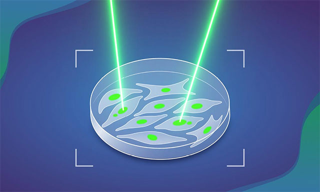
EMBL's AI MAGIC uses molecular laser tagging technology to mark and analyze cells with chromosomal abnormalities, revealing the first steps in the cancer development process.
Table of Contents
Researchers at EMBL have developed a new AI tool that uses 'molecular laser tagging' to identify cells that may reveal the earliest origins of cancer.
The human body relies on precise genetic instructions for cells to function properly. Cancer begins to form when these instructions become disrupted. As genetic errors accumulate over time, cells can lose control of their growth and begin multiplying uncontrollably. Among the earliest changes that lead healthy cells to the precancerous stage are chromosomal abnormalities – including errors in number or structure.
At EMBL Heidelberg, Korbel's research team created an AI-based tool that allows scientists to closely observe how these abnormalities appear. The insights gained from this method could help shed light on the initial steps in the cancer development process.
" Chromosomal abnormalities are a leading factor in particularly aggressive forms of cancer, and are strongly linked to mortality, metastasis, recurrence, chemotherapy resistance, and tumor growth rate ," shared scientist Jan Korbel, lead author of the work published in Nature. " We wanted to understand what determines a cell's susceptibility to these changes, and how quickly they occur when a healthy cell replicates ."
The link between chromosomal abnormalities and cancer was actually established more than a century ago, when scientist Theodor Boveri observed under a microscope and hypothesized that uneven chromosomal content could promote cancer formation.
Why are chromosomal abnormalities so difficult to study?
Although known for a long time, detecting these cells remains extremely difficult because they are very rare, and many of them die or are eliminated by the body's natural mechanisms. Researchers often have to search manually under microscopes and only manage to collect a small number of cells for further analysis.
Scientist Marco Cosenza from Korbel's group found a solution after collaborating with several groups at EMBL facing similar problems. He and his colleagues developed an automated system combining automated microscopy, single-cell sequencing, and AI. They called it MAGIC – an acronym for machine learning-assisted genomics and imaging convergence .
Molecular 'laser tagging' for cell identification and labeling.
Essentially, MAGIC operates like a fully automated 'laser gun': it searches for 'objects' exhibiting specific characteristics. In this study, the scientists focused on a cellular structure called a micronucleus – tiny compartments containing fragments of DNA that are separated from the majority of the genome. Cells with micronucleus often exhibit more chromosomal abnormalities, making them more susceptible to cancer.
When a cell containing micronucleus is detected, the system 'shoots a laser beam' to label it. Specifically, they use a dye that changes color and glows when exposed to light, allowing for permanent cell marking.
Cosenza said the project combines many of his interests: from genomics and microscopy to robotic automation. During the COVID-19 lockdown in 2020, he learned and applied AI technology to process biological images from previous data, then designed experiments to test and improve it.
The MAGIC process works as follows: an automated microscope captures a series of images of cell samples; a machine learning algorithm, trained on a manually labeled set of images, scans and detects cells with micronucleus; the cell's location is sent back to the microscope for illumination and labeling. Labeled cells can then be easily separated using techniques such as flow cytometry for further analysis, e.g., genome sequencing.
Scaling up a process that is inherently very slow.
Automating a manual, slow, and error-prone process has allowed MAGIC to accelerate research to unprecedented levels: in less than a day, it can analyze nearly 100,000 cells.
The research team used MAGIC to analyze cell cultures derived from normal human cells. The results showed that over 10% of cell divisions exhibited spontaneous chromosomal abnormalities, and this number nearly doubled when the p53 gene – a well-known tumor suppressor gene – was mutated. They also investigated other factors such as the location and presence of double-strand DNA breaks.
The research involved collaboration between several groups within and outside EMBL, including the Advanced Light Microscopy Facility (ALMF), the Pepperkok group at EMBL Heidelberg, Isidro Cortes-Ciriano's group at EMBL-EBI, and Andreas Kulozik's group at the German Cancer Research Centre.
MAGIC is a flexible technique that can be trained in many ways. Although this research focuses on micronucleus, the algorithm could theoretically be trained to detect many other cellular features.
"If that feature is distinguishable to the naked eye from a normal cell, AI can learn to recognize it ," Korbel said. " This system has the potential to drive many new discoveries in biology ."
Was this article helpful?
Your feedback helps us improve.
Related Articles
 7 symptoms of early cancer are often neglected by 90% of people6 minutes read
 FDA unexpectedly approved Nerlynx therapy to reduce the risk of breast cancer recurrence2 minutes read
 Protein analysis in breast milk helps detect breast cancer early2 minutes read
 Recognize early signs of throat cancer for prompt treatment4 minutes read
 Why are people who take advantage of the morning more successful?9 minutes read
 Successful manufacture of new medical ink marks the goal of radiotherapy to treat skin cancer2 minutes read
Reader Comments 0
Sign in with email or Google to join the discussion.