It took 1700 hours to describe the brain of 'fruit flies' in 3D
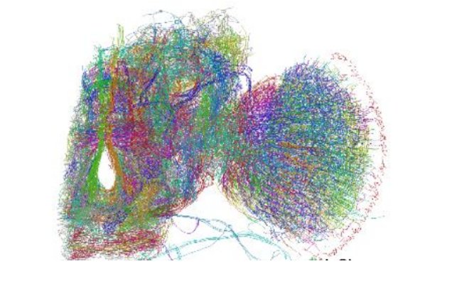
Recently, a group of Tokai University scientists in Japan introduced the first three-dimensional ( 3D ) model of the " network " of Neonal network of fruit flies . The study of this group of scientists is an important step forward for neuroscience ( neuroscience insofa ) because it overcomes limitations in previous brain imaging techniques by giving an image. High resolution Neuron network, capable of describing the shape and location of about 100,000 neurons.
 Photo source: Mizutani et al / Arxiv.
As detailed in an article published on arxiv last month, researchers were able to accomplish this by re-setting the usual technical image target to create tissues. 3D shapes of complex molecules.
Known as an X -ray crystallography ( x-ray crystallography - the science of determining the arrangement of atoms within a crystal based on data on the dispersion of X-rays after projecting into the crystalline electrons ), this technique works with the first pure crystalline molecular molecule being studied. After constructing 3D images of X-rays and X-ray diffraction patterns, the results were obtained. The problem here is that the results of the X-ray diffraction model actually only show the density of the electrons inside the crystal, not the position of the average atom - which is indicated by the diffraction measurements. electron.

However, when the Tokai team discovered and utilized this technique to describe a neural network (" networks of nerves" ) was a bit complicated. This is because atoms in the same molecule are different - they are considered as points lying in space. Neurons in the brain are like winding bends, making it difficult for researchers to illustrate their position and structure based on diffraction data.
Therefore, the team gradually refined this approach to images by using a technique called X -ray tomography ( x-ray tomography ). Instead of creating a brain- crystallized version of the fruit fly, the team immersed their brains in a silver dye solution. They then put the silver-dyed brain into X-rays to measure how much silver in the nerve cells absorbed the radiation. The data given by this technique is included in a computer imaging program, which uses that data to create a 3D model of the shape and location of nerve cells in the brain. of fruit flies.
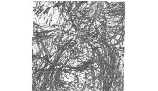
 The fruit fly's neuron network was captured at a resolution of 140 nanometers.Photo source: arXiv / Mizutani et al.
The final result of the goal of this biological imaging technique describes the neuron network of fruit flies as impressive. The first 3D image shows the location and nerve connections at a resolution of about 600 nm ( nanometer ), describing about 100,000 neurons. In particular, most of these neurons are represented by computer models, which can check the appropriateness of nerve connections. In addition, when any abnormality occurs, scientists can correct errors by hand.
By the time everything was completed and published, the researchers had to spend 1700 hours to create a " neural " network ( neuron ) - nearly 71 days round. This is a huge investment to be able to apply to neuron networks in the human brain: about 100 billion neurons. Obviously, this technique is not usable for the human brain however, it is an important step to figure out how to illustrate more complex brains.
You should read it
- ★ You are often teased as the 'goldfish brain', do not be sad this indicates you have a brain that works very well
- ★ How to download Brain Out on the computer, install Brain Out on the computer
- ★ Interesting discovery: Human brain is more flexible than chimp brain
- ★ Stem cells help patch brain damage in stroke victims
- ★ Answers to Test Brain Level 1 to 60 (updated continuously)